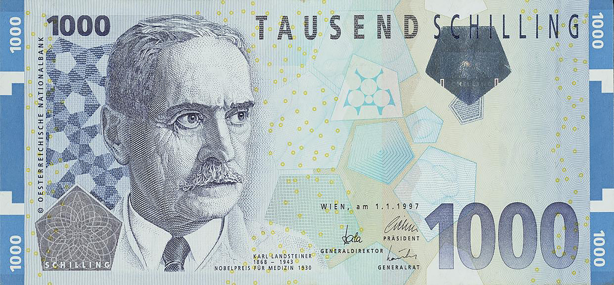
| ABO discovered | Karl Landsteiner, 1901 (Nobel Prize, 1930) |
|---|---|
| AB group added | 1902, by von Decastello and Sturli |
| Rh factor | 1940, by Landsteiner and Wiener (named for the Rhesus macaque) |
| ABO types | A, B, AB, O |
| Universal donor | O negative (red cells) |
| Universal recipient | AB positive (red cells) |
| Global population | O+ (39%), A+ (27%), B+ (22%), O− (3%), A− (2%), AB+ (2%), B− (1%), AB− (1%) |
| (population-weighted world estimate) | |
What a Blood Group Is
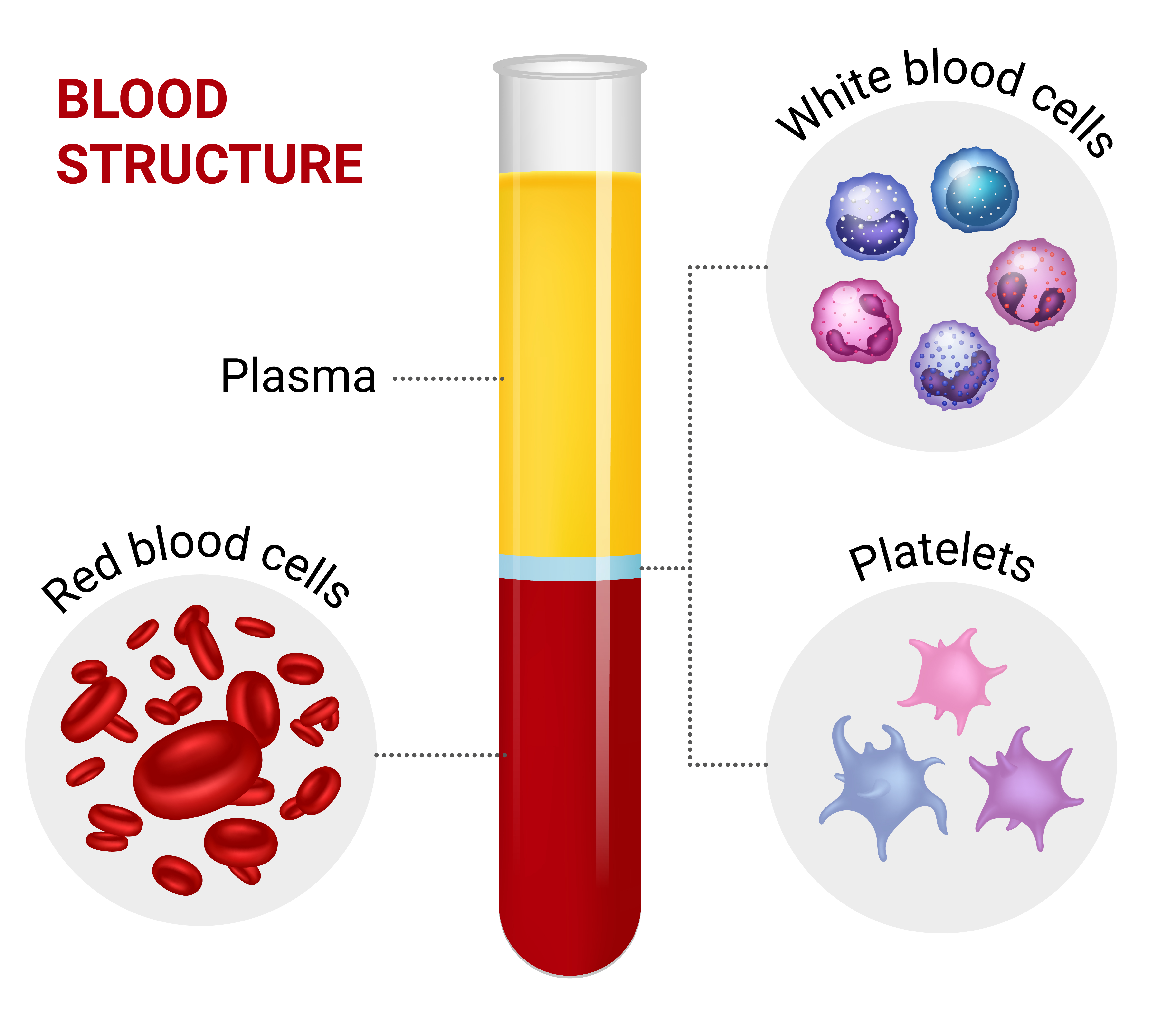
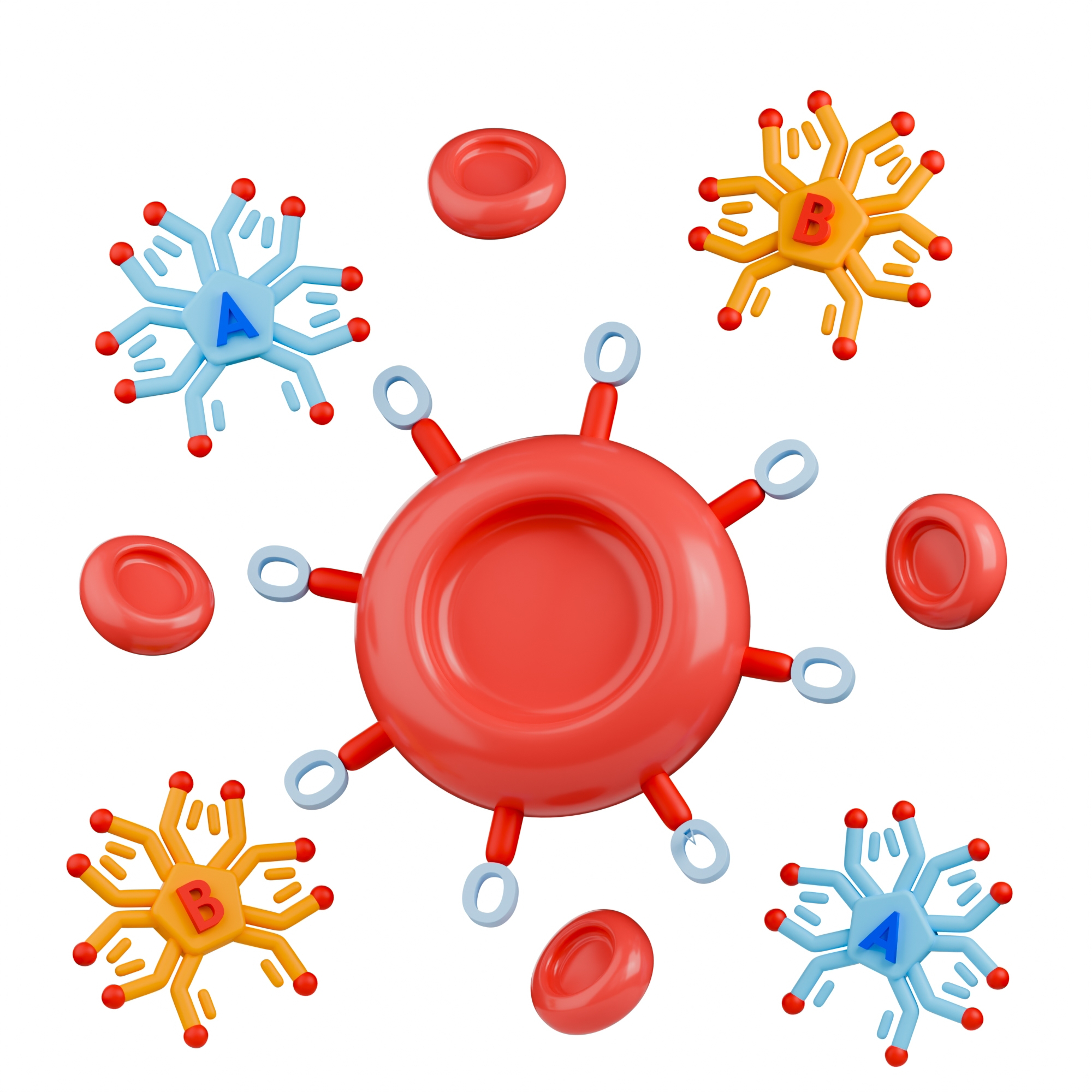
A blood group is a way of classifying blood by the inherited markers on the surface of its red blood cells. These markers, called antigens, are proteins or sugars that stud the cell membrane. The immune system treats them as identity badges: it tolerates the antigens the body was born with and attacks any it does not recognize.
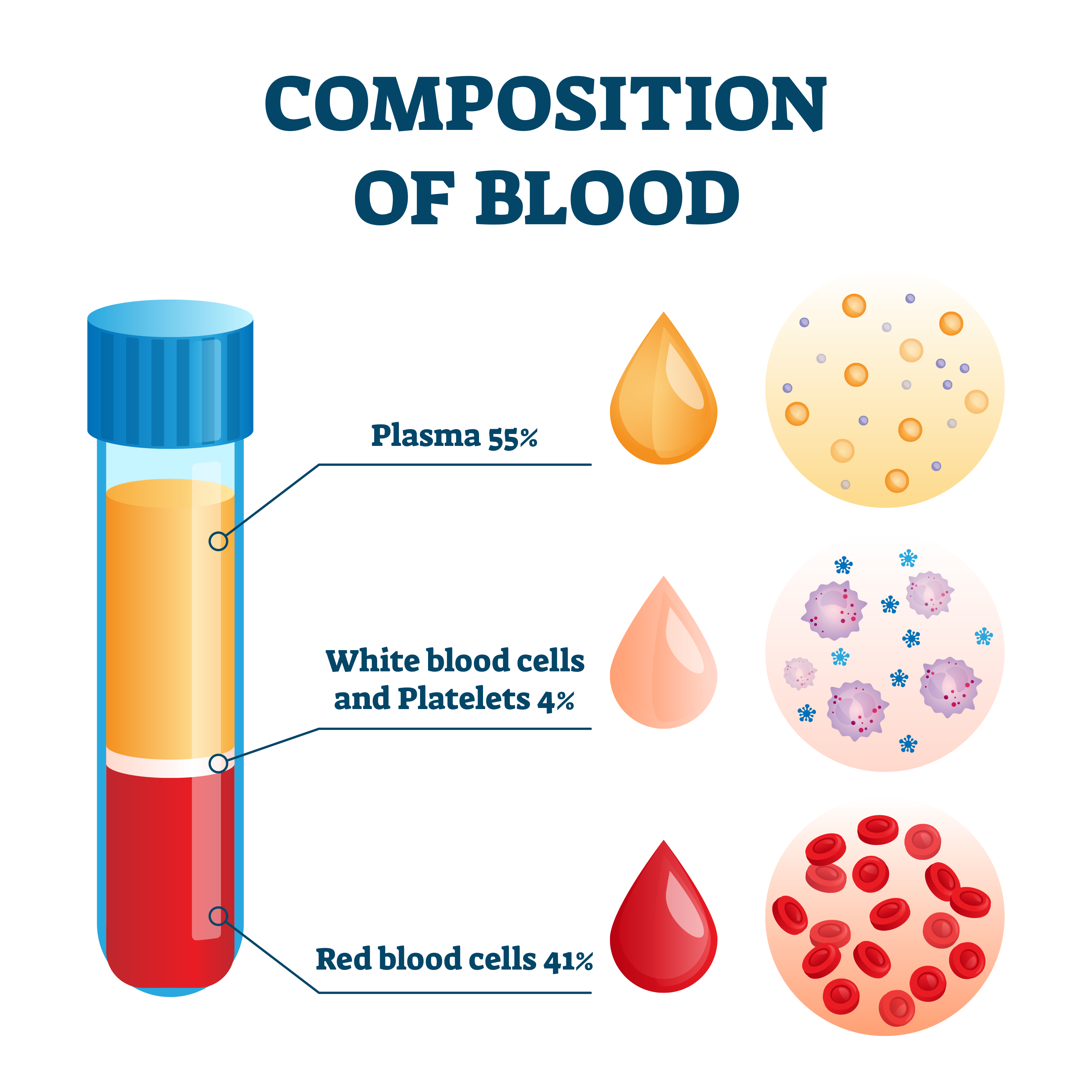
The reason this matters is defensive chemistry. Floating in the plasma are antibodies, immune proteins shaped to lock onto foreign antigens. When an antibody meets its matching antigen on a red cell, it bridges cells together and they clump — a reaction called agglutination. In a test tube that clumping is how blood is typed. In a vein it is a medical emergency, because clumped and ruptured cells can block vessels, starve organs, and trigger shock.
Blood group antigens are inherited, following ordinary rules of genetics, which is why blood type runs in families and why it can settle questions of parentage. And while the ABO and Rh systems dominate everyday medicine, they are only the beginning: the International Society of Blood Transfusion recognizes 48 distinct blood group systems, together carrying several hundred red-cell antigens; the two newest, MAL and PIGZ, were added as systems 47 and 48. Most never trouble a patient. A handful can be as dangerous as ABO.
The ABO System
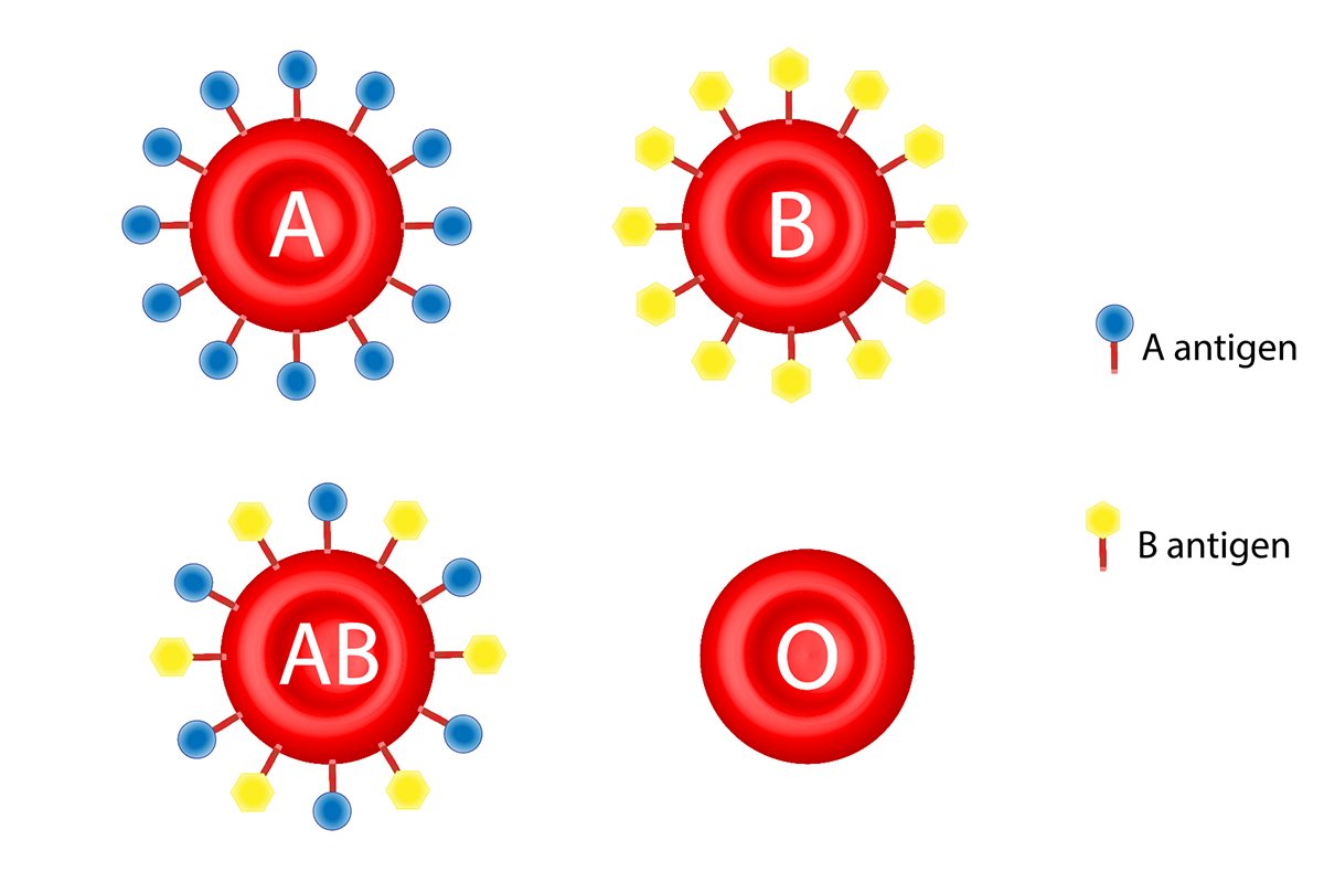
The ABO system is the foundation of transfusion medicine, and it works on a strict logic of matching antigen to antibody. When Landsteiner sorted his 1901 samples, he named each group for the antigen its red cells carried.
He found three. He called the first two A and B, after the A and B antigens on their red cells. The third group’s cells carried neither antigen, so he labeled it C. He soon renamed that third group O — from the German ohne, meaning “without” — a reminder that its cells carry no A or B antigen at all. The fourth and rarest group, AB, whose cells carry both antigens, was identified the following year, in 1902, by his colleagues Alfred von Decastello and Adriano Sturli.
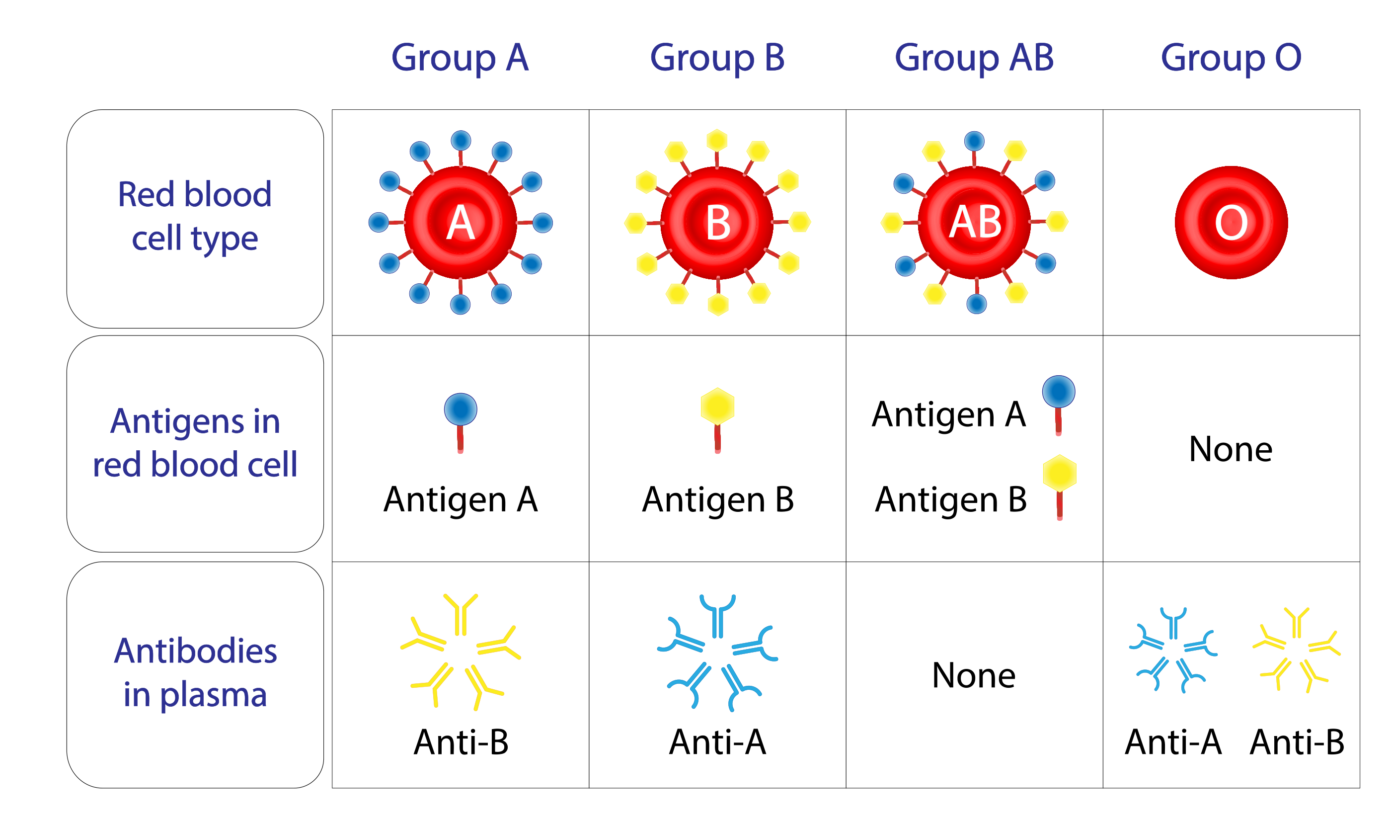
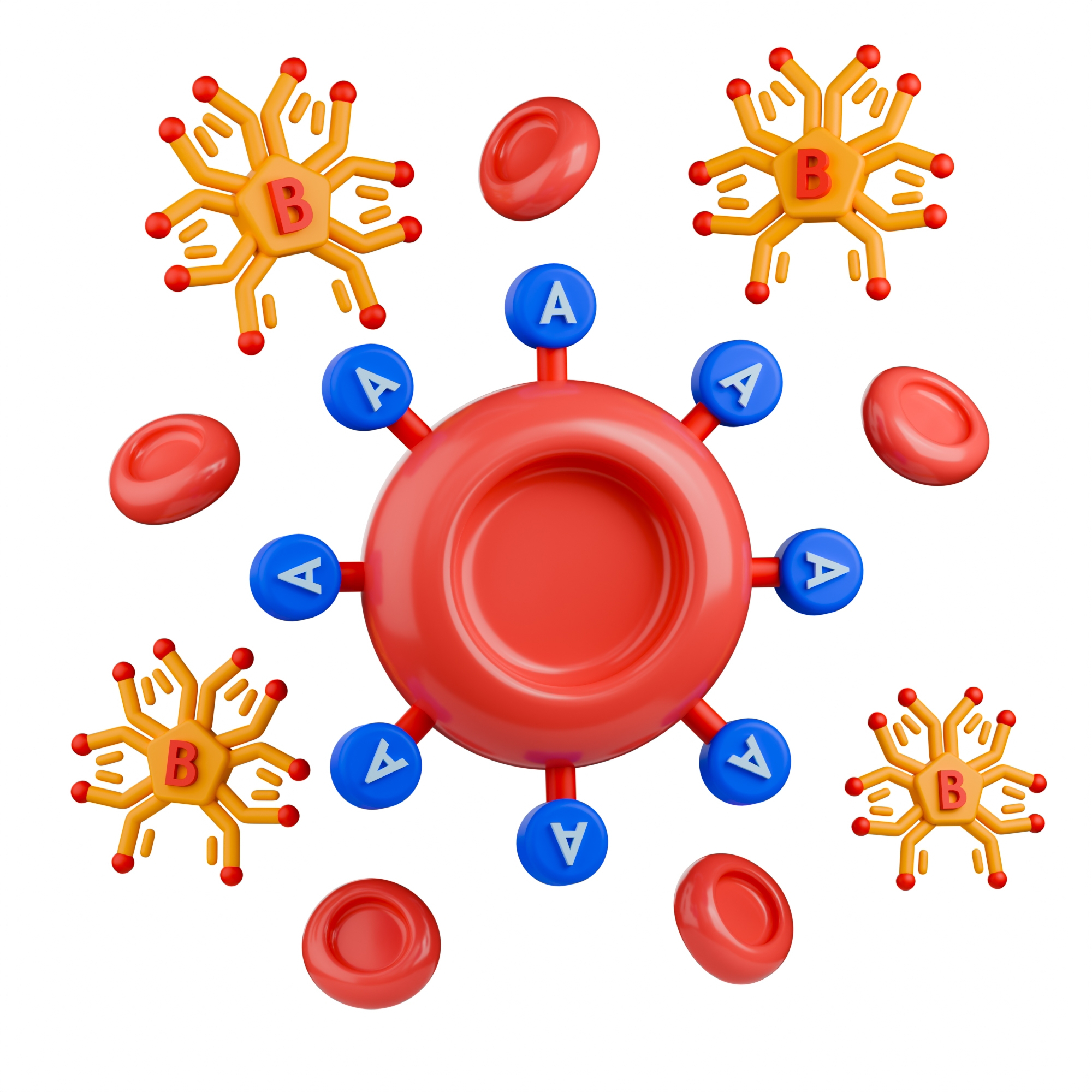
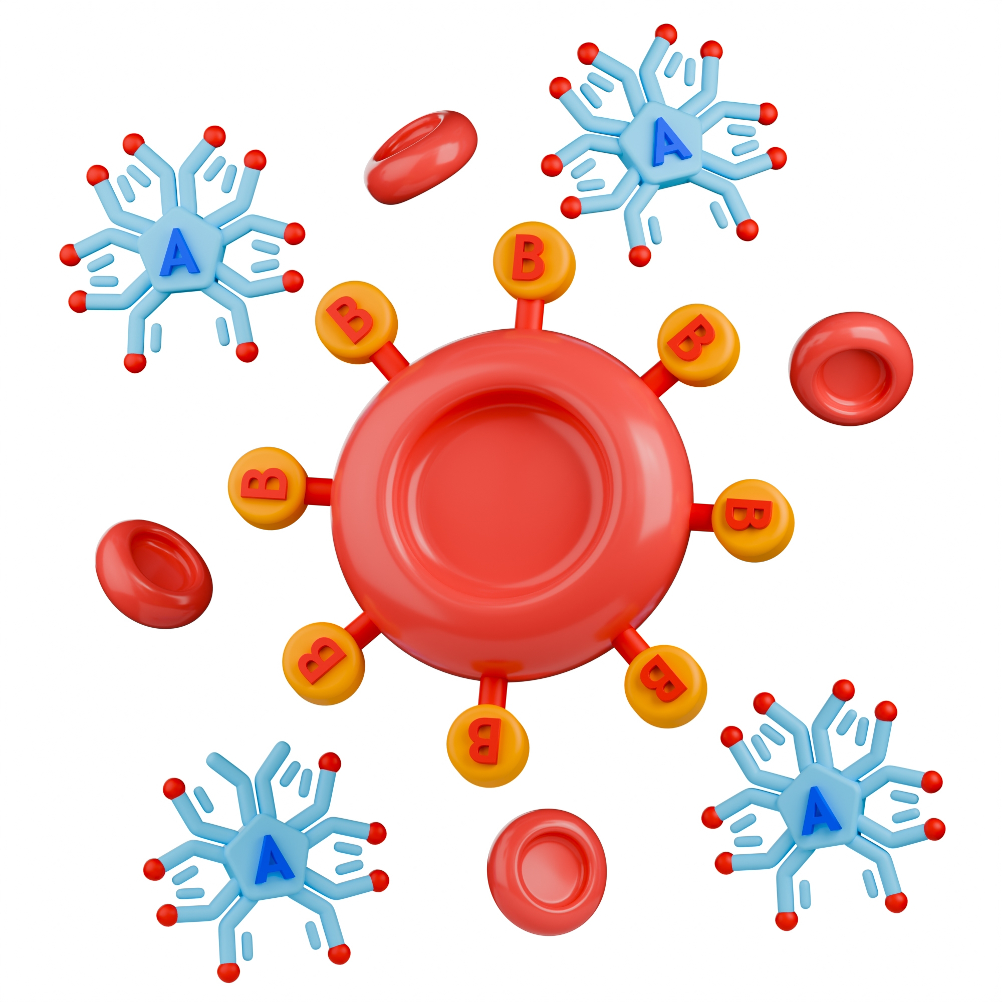
Every ABO type is defined by two things at once: which antigens sit on the red cells, and which antibodies float in the plasma. The pairing is always opposite — the body makes antibodies against the antigens it lacks. These antibodies are “naturally occurring”: they appear in early childhood, without any transfusion, prompted by exposure to common environmental substances that resemble the A and B antigens.
| ABO type | Antigens on red cells | Antibodies in plasma |
|---|---|---|
| Type A | A antigen | anti-B |
| Type B | B antigen | anti-A |
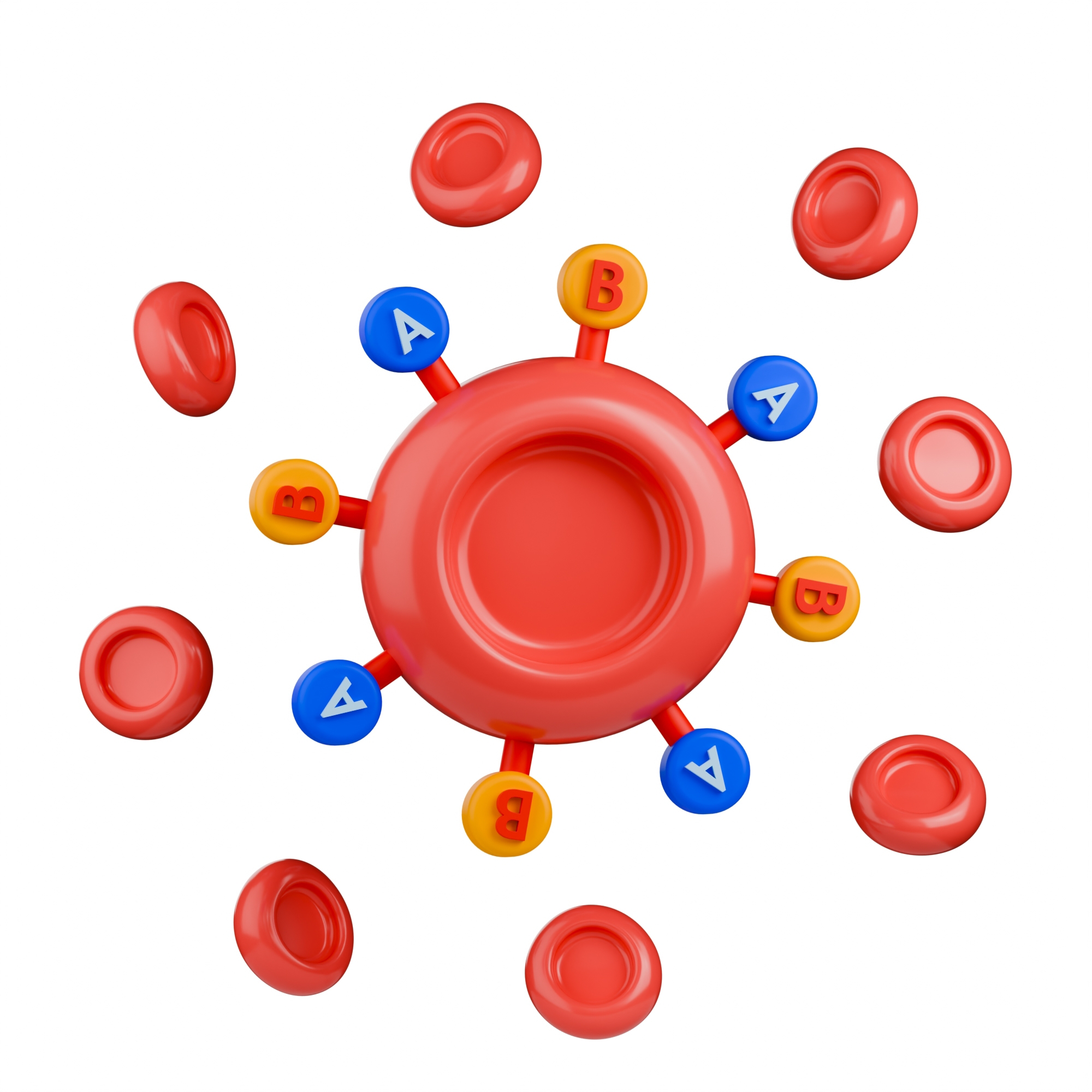
| Type AB | A and B antigens | neither anti-A nor anti-B |
| Type O | neither A nor B antigen | both anti-A and anti-B |
Those two markers set up a neat opposition at the extremes. Type AB wears both antigens and makes no ABO antibodies at all, which is why it can accept red cells of any ABO type. Type O, wearing no antigens but armed with both antibodies, is the mirror image: its cells offend no one, but its plasma attacks almost everyone.
The H Antigen Beneath A and B
A and B share a hidden foundation. Both are built by adding a sugar onto a base molecule called the H antigen, produced by a gene called FUT1. An A gene adds one kind of sugar to H; a B gene adds a different one; the common O gene adds nothing, leaving H bare. So type O cells are not chemically blank — they are covered in unmodified H antigen. That detail sounds academic until it explains one of the rarest blood types on Earth, the Bombay phenotype, described later.
The Rh Factor
Nearly four decades after ABO, a second system emerged that would prove just as consequential, especially for mothers and newborns. In 1940, Karl Landsteiner and Alexander Wiener described the Rh factor after immunizing rabbits and guinea pigs with the red cells of the Rhesus macaque monkey (Macaca mulatta) — the animal that gave the system its two-letter name. Later work showed the human D antigen was not identical to the original monkey-associated one, but the “Rh” name had already stuck. So despite the name, Rh-positive people carry no “monkey protein” — only human Rh proteins made by human genes.
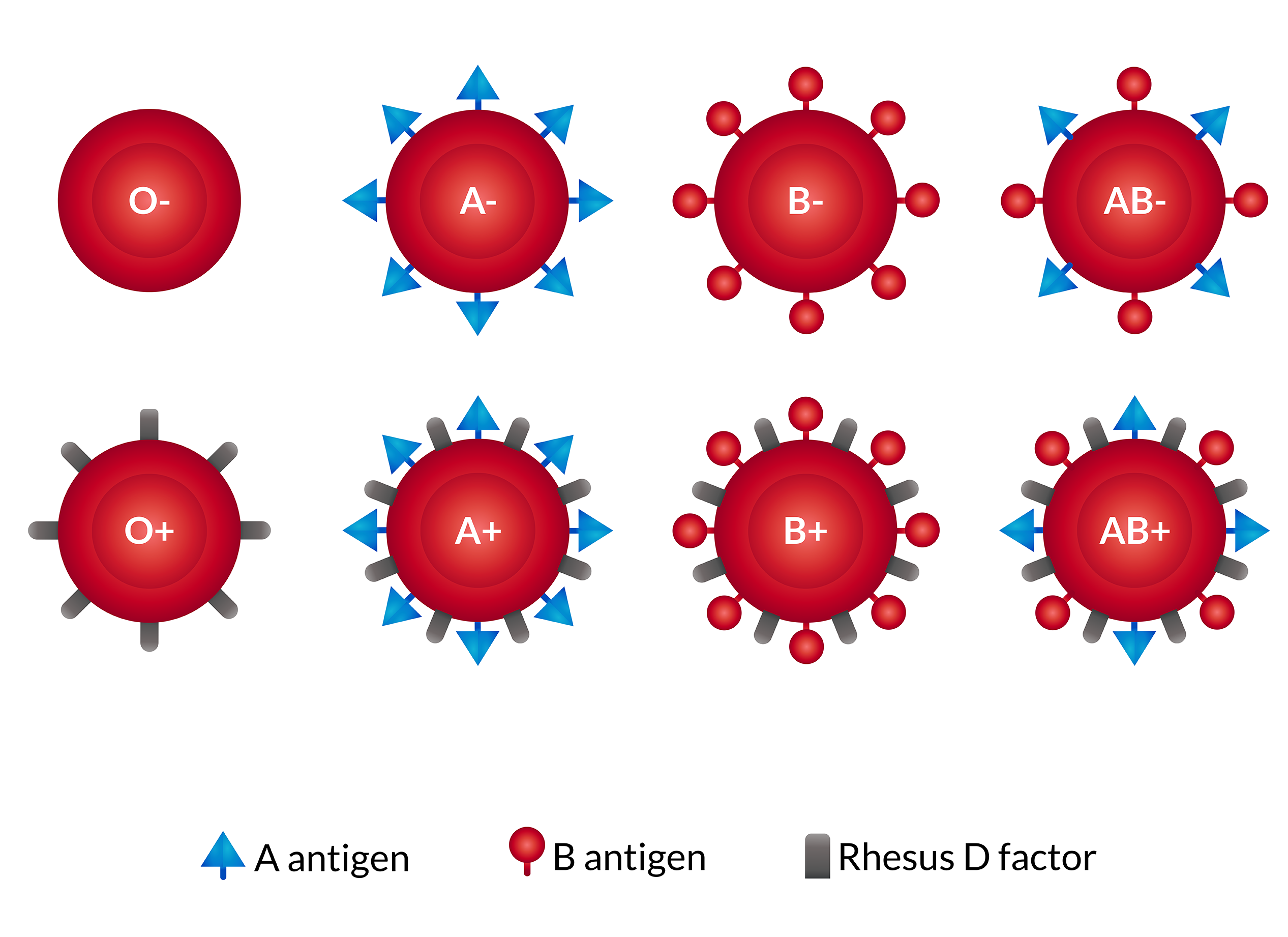
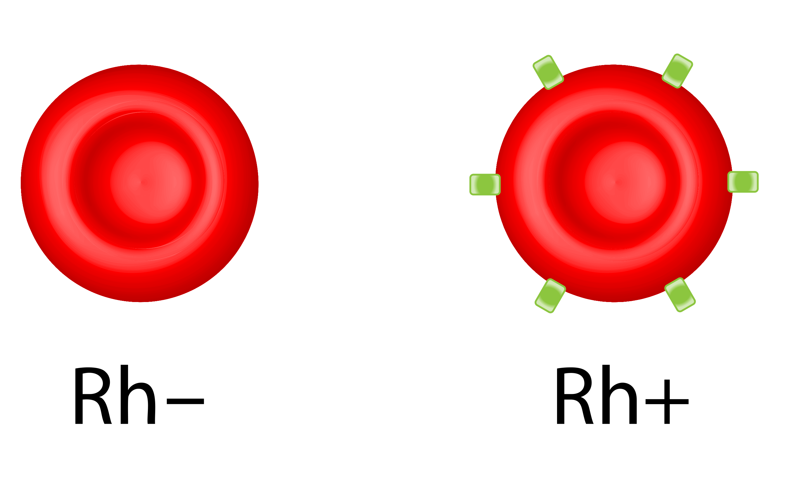
The Rh system is large and complex — the ISBT’s May 2025 table listed 56 Rh antigens — but one dominates: the D antigen. In everyday language, having the D antigen makes a person “Rh-positive” and lacking it makes them “Rh-negative.” That single plus or minus, appended to the ABO letter, produces the familiar eight-way labels from O-negative to AB-positive. The popular phrase “Rh factor” is convenient but slightly misleading: it makes the system sound like one substance, when the plus or minus really reports just the D antigen out of dozens.
| Rh status | Meaning |
|---|---|
| Rh-positive (+) | red cells carry the D antigen |
| Rh-negative (−) | red cells lack the D antigen |
The Rh factor behaves differently from ABO in one critical way. A person does not make anti-D antibodies naturally; they make them only after being exposed to Rh-positive blood — through a mismatched transfusion or, as the next sections show, through pregnancy. That delayed reaction is what makes Rh both manageable and, if ignored, dangerous.
Transfusion Compatibility
The governing rule of transfusion is blunt: a person cannot receive an antigen their body does not already possess. Introduce a foreign antigen and the recipient’s waiting antibodies attack the donated cells, causing an acute hemolytic transfusion reaction — the immune destruction of the transfused blood, which can be fatal. Safe transfusion therefore matches both the ABO group and the Rh factor.
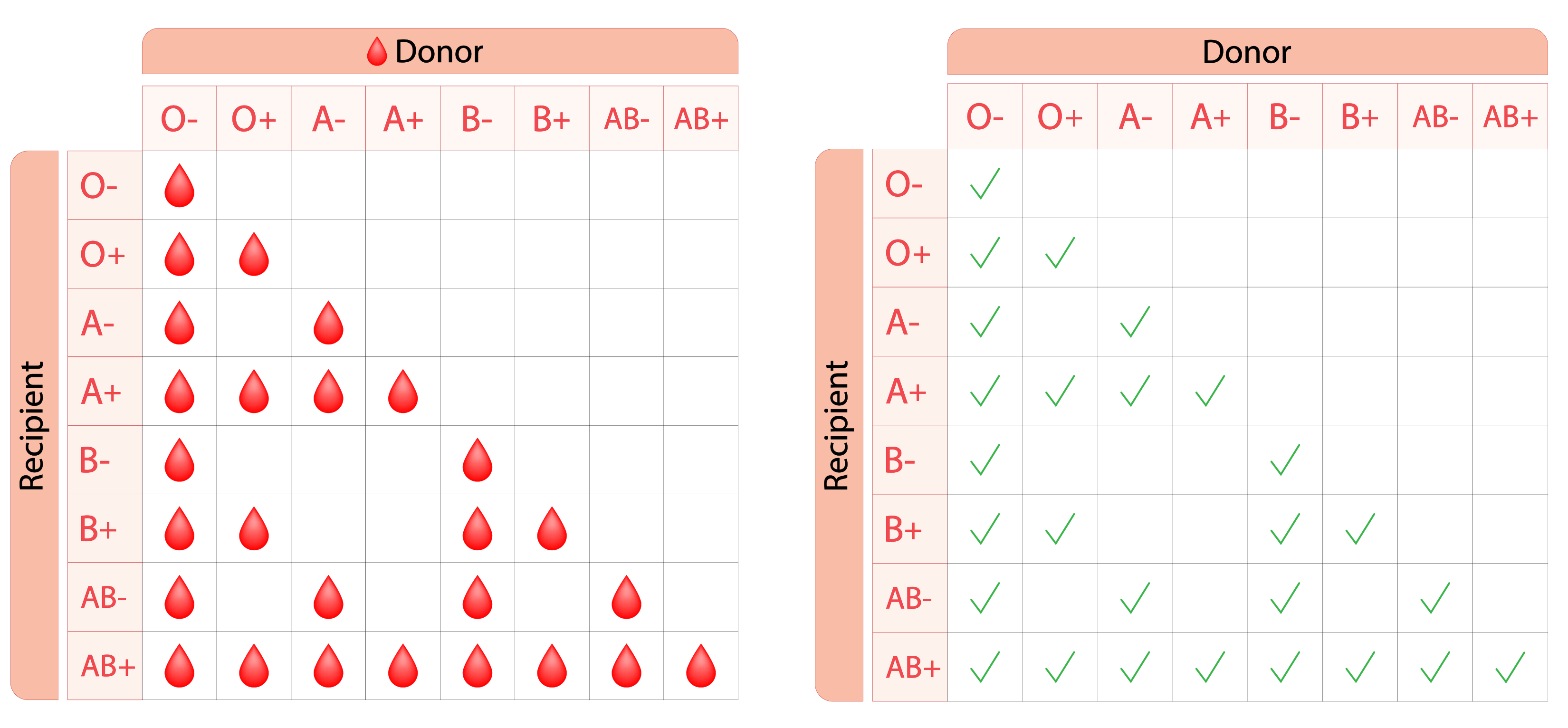
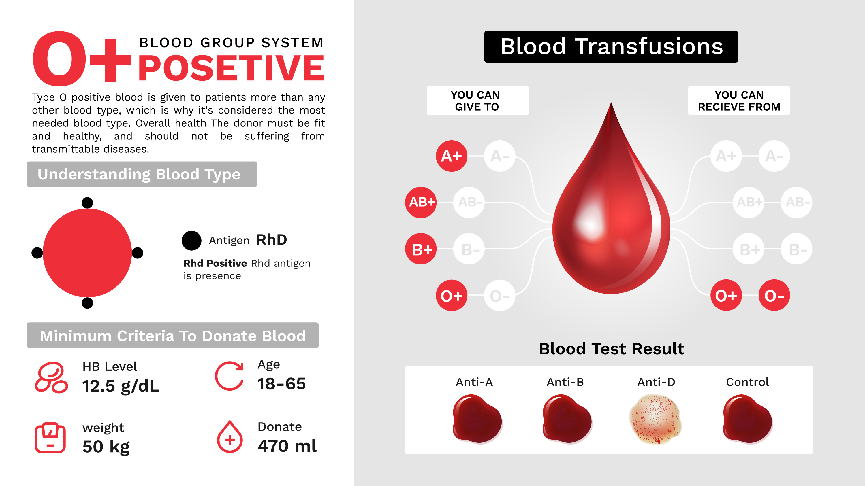
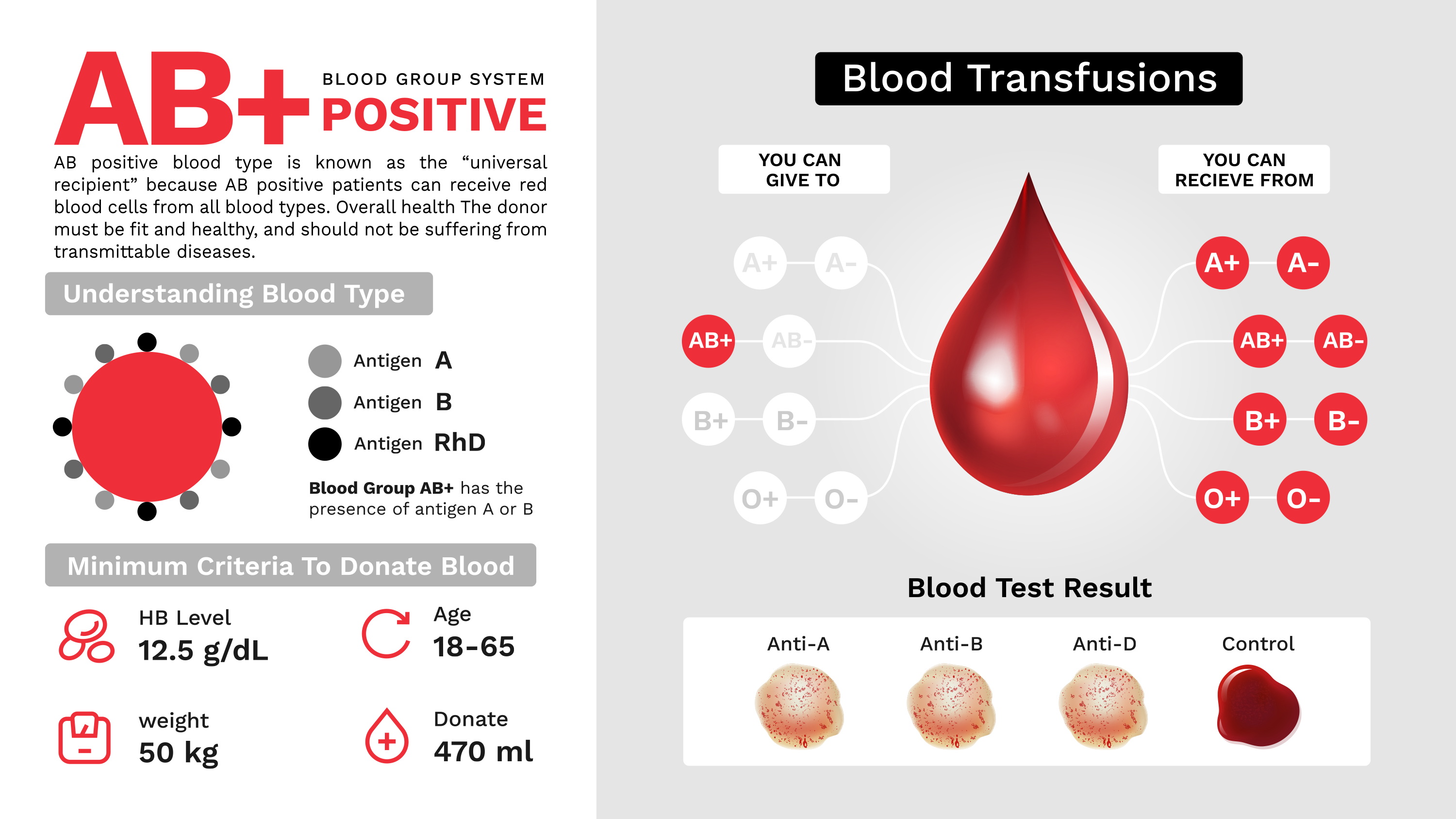
Two types anchor the extremes. Because O-negative cells carry no A, no B, and no D antigen, the immune system finds nothing foreign to attack, making O-negative the universal red-cell donor and the default in an emergency when there is no time to type a patient. Because AB-positive people already carry every one of those markers, their immune system recognizes all of them, making AB-positive the universal red-cell recipient.
| Blood type | Can receive red cells from | Can donate red cells to |
|---|---|---|
| O− | O− | all eight types (universal donor) |
| O+ | O−, O+ | O+, A+, B+, AB+ |
| A− | O−, A− | A−, A+, AB−, AB+ |
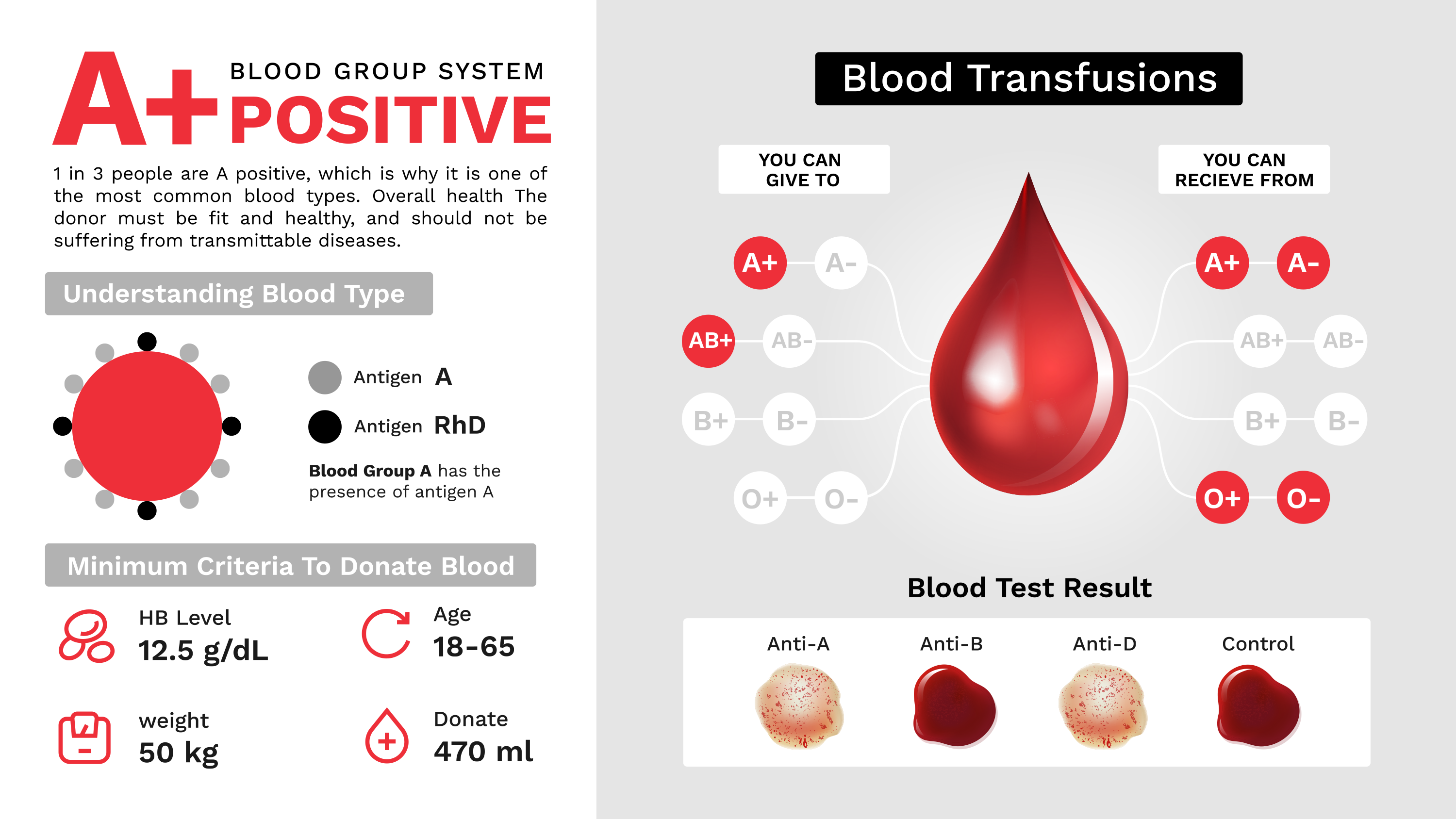
| A+ | O−, O+, A−, A+ | A+, AB+ |
| B− | O−, B− | B−, B+, AB−, AB+ |
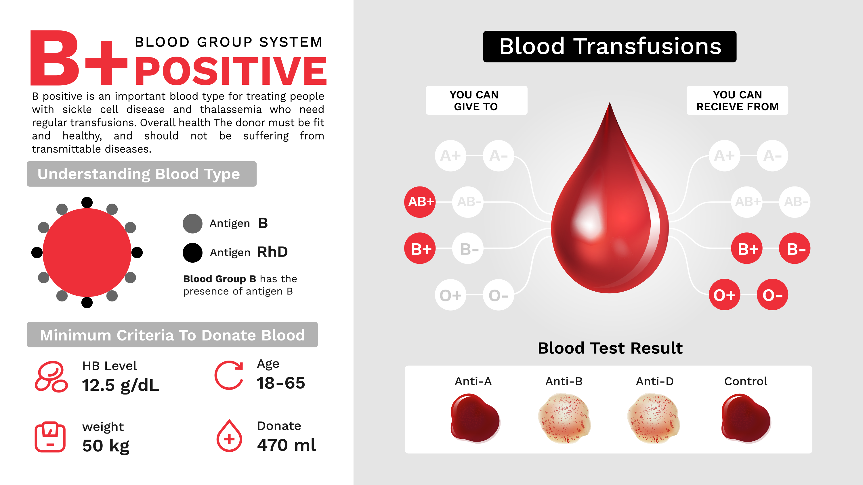
| B+ | O−, O+, B−, B+ | B+, AB+ |
| AB− | O−, A−, B−, AB− | AB−, AB+ |
| AB+ | all eight types (universal recipient) | AB+ |
One twist catches many people out: for plasma, the rules invert. Plasma carries antibodies rather than antigens, so the safe donor is the one whose plasma contains no ABO antibodies to attack the recipient’s cells. That makes AB the universal plasma donor and O the universal plasma recipient — the exact reverse of the red-cell picture. It is a favorite examination trap, and the reason the phrase “universal donor” always needs the words “red cell” or “plasma” attached.
The ABO gene is older than our species — it has run in primate blood for more than 20 million years.
Inheritance and Paternity
Blood type is a genetic trait, passed from parents to children by clear rules. In the ABO system, the A and B genes are co-dominant — if a child inherits one of each, both are expressed, producing type AB — while the O gene is recessive, showing up only when a child inherits it from both parents. For the Rh factor, positive is dominant and negative is recessive, so an Rh-negative child must have inherited a negative gene from each parent.
These rules make blood typing a tool in questions of parentage. It cannot prove that a particular man is a child’s father, but it can prove that he is not, because a child cannot carry an ABO antigen that neither parent possesses. If a child is type AB and the mother is type O, for instance, the father must have supplied either an A or a B gene — a man who is type O could not have.
| Parent 1 | Parent 2 | Possible children | Impossible |
|---|---|---|---|
| O | O | O | A, B, AB |
| A | O | A, O | B, AB |
| B | O | B, O | A, AB |
| A | B | A, B, AB, O | none |
| AB | O | A, B | O, AB |
| AB | AB | A, B, AB | O |
The AB × O row holds a genuine curiosity: two parents can produce children whose blood types match neither of them, because an AB parent passes on either A or B (never both to one child), and the O parent passes on O, yielding type A or type B offspring.
Rh Incompatibility in Pregnancy
The most far-reaching consequence of the Rh factor plays out before birth. The danger arises in one specific situation: an Rh-negative mother carrying an Rh-positive baby, a combination possible when the father is Rh-positive.
The mechanism unfolds across two pregnancies. During delivery — or any event that mixes fetal and maternal blood — some of the baby’s Rh-positive cells can cross into the mother’s bloodstream. Her immune system, meeting the D antigen for the first time, begins manufacturing anti-D antibodies. A first Rh-positive baby is usually unharmed, because the antibodies form too late to affect it. But in a subsequent pregnancy those antibodies can cross the placenta and attack the red cells of another Rh-positive fetus, destroying them. The result is hemolytic disease of the newborn, also called erythroblastosis fetalis, which can be fatal to the fetus or newborn.
The solution is one of modern medicine’s quiet triumphs. Rh-negative mothers are given injections of Rh immunoglobulin — sold as RhoGAM and similar brands — typically around week 28 of pregnancy and again within 72 hours of delivery. The injected antibodies clear the fetal Rh-positive cells from the mother’s circulation before her own immune system can respond, so she never makes the dangerous anti-D antibodies, and future pregnancies stay safe.
The textbook version does oversimplify one thing. Anti-D is the most famous cause of hemolytic disease of the newborn, but not the only one: antibodies to other red-cell antigens — c and K (Kell) among them — can cause it too, and ABO incompatibility produces a usually milder form. The risk also depends on the mother actually becoming sensitized, not merely on a mismatch in type. The neat rule “Rh-negative mother plus Rh-positive baby equals danger” captures the main anti-D story but hides these other paths.
Minor Systems and the Rarest Blood
ABO and Rh dominate the clinic, but they share the red cell’s surface with dozens of other systems, some of them medically serious and one of them almost mythically rare.
Three minor systems earn regular attention. The Kell system carries a highly immunogenic K antigen, meaning it provokes strong antibodies that can cause severe transfusion reactions and fetal anemia. The Duffy system is famous beyond hematology: certain Duffy antigens act as the doorway that the malaria parasite Plasmodium vivax uses to enter red cells, and many people of African descent lack these antigens. That absence strongly reduces susceptibility to vivax malaria — though the accurate word is “resistance,” not “immunity,” since infections in Duffy-negative people have been documented. The Kidd system is known for a dangerous stealth — its antibodies can fade below detectable levels between exposures, then surge on re-exposure, causing delayed transfusion reactions. Beyond these, systems such as MNS and Diego matter in transfusion and, because their antigen frequencies vary sharply by region, in tracing human populations.
There is also a type that hides in plain sight: the Bombay phenotype, first described in Bombay (now Mumbai) in 1952. These rare individuals inherit no working H antigen at all, so — recalling that A and B are built on H — they can build neither A nor B even if they carry those genes. On a routine test they look like type O. But their plasma contains an extra antibody, anti-H, and because ordinary O cells are covered in H antigen, a Bombay patient given normal O blood can suffer a severe reaction. They can safely receive blood only from another Bombay donor, which is why rare-donor registries exist. In India, where it was first found, the phenotype occurs in roughly one person in 10,000; elsewhere it is far rarer.
The rarest blood of all is called “golden blood,” known technically as Rh-null: blood whose cells lack every antigen in the entire Rh system, not just the D. Canadian Blood Services reports that fewer than 50 people worldwide have ever been identified with it — a figure best read as an attributed estimate rather than a live census. Golden blood is priceless for transfusion research because, lacking all Rh antigens, it can in principle be given to patients with rare Rh types who can accept nothing else. Yet it is perilous for the people who carry it, because they in turn can accept blood from no one but another Rh-null donor — a population small enough to fit in a single room.
The Blood of Other Animals

One of the most surprising facts about human blood groups is that they are not entirely human. The ABO antigens are far older than our species, and versions of the same system run in the veins of our closest relatives.
Among the great apes, the resemblance is striking. Chimpanzees and bonobos have overwhelmingly type A blood, with a smaller share of chimpanzees carrying type O. Orangutans display all four ABO types — A, B, AB, and O. Gorillas are the outliers: they do not fit neatly into the human ABO scheme, and while older studies described a “B-like” antigen, molecular work suggests the gorilla’s blood groups diverge from the human system. All the great apes can also be Rh-positive or Rh-negative, though their D antigens differ slightly from ours. Old World monkeys, and even some baboons, carry recognizable ABO substances too.
Chimpanzees are almost all type A — yet no ape can safely give blood to a human of the “same” type.
How deep does the kinship run? The ABO gene traces back to a common primate ancestor that lived more than 20 million years ago, and blood group A is thought to be the oldest of the ABO types in primates. A landmark 2012 study went further, arguing that the A and B variants form a “trans-species polymorphism” — meaning humans share the very same ancient A and B gene variants with other primates, inherited unbroken across millions of years rather than re-evolved separately. (An earlier analysis had suggested chimpanzee A and gorilla B arose by convergent evolution; the trans-species picture is now the stronger view for the A/B alleles.)
The kinship comes with a hard limit, and it makes a perfect quiz answer: despite sharing the ABO labels, apes and humans cannot safely swap blood. A type A chimpanzee cannot donate to a type A human, nor the reverse — millions of years of subtle molecular drift have made the “same” blood types incompatible across species. The shared alphabet is real; the blood behind it is not interchangeable.
Discovery: A History Written in Blood
The science of blood groups grew out of centuries of dangerous trial and error. The essential groundwork was laid in 1616, when the English physician William Harvey announced that blood circulates through the body in a closed system, publishing his landmark treatise on the motion of the heart and blood in 1628 — a prerequisite for the very idea of transfusion.
Experiments followed, and they were grim. In 1665 the physician Richard Lower performed transfusions between dogs in England, and in November 1667 he transfused lamb’s blood into a man. In France, Jean-Baptiste Denis, physician to King Louis XIV, was transfusing lamb’s blood into human subjects and recorded what is likely the first account of a hemolytic transfusion reaction. After a patient died, Denis was arrested, and in 1668 the transfusion of animal blood into humans was banned in France; the British Parliament followed with its own prohibition in 1678. For roughly 150 years, the field barely moved.
Progress resumed in the 19th century. The English obstetrician James Blundell, appalled by mothers bleeding to death after childbirth, argued that human patients should receive human blood, and performed transfusions on that principle. In 1875 the German physiologist Leonard Landois showed scientifically why cross-species transfusion failed: mixing one species’ red cells with another’s serum made the cells clump and burst. The stage was set for Landsteiner’s 1901 breakthrough, which finally explained why even human-to-human transfusion so often went wrong — and turned a deadly gamble into a routine, life-saving procedure.
Discovery did not stop with the four groups. In 1907 Reuben Ottenberg put compatibility testing — the crossmatch — into transfusion practice, cutting fatal mismatches. In 1914 sodium citrate was introduced to stop donated blood from clotting, so it no longer had to flow vein-to-vein and could be stored, making blood banks possible. In 1939 Philip Levine and Rufus Stetson traced a mother’s severe reaction and fetal loss to a red-cell antibody — the first glimpse of what became Rh disease, a year before the Rh factor was named. And in 1945 Robin Coombs, Arthur Mourant, and Rob Race introduced the antiglobulin (Coombs) test, which finally exposed the “hidden” antibodies that coat red cells without clumping them — the tool that made modern antibody screening, and much of the rest of this story, possible.
Timeline / Milestones
- 1616
Harvey describes circulation
William Harvey shows blood moves in a closed loop, a prerequisite for transfusion (published 1628).
- 1665
First animal transfusions
Richard Lower transfuses blood between dogs; in 1667 he gives a man lamb's blood.
- 1668
Animal-to-human transfusion banned
After a death under Jean-Baptiste Denis, France bans it; Britain follows in 1678.
- 1901
ABO system discovered
Karl Landsteiner identifies three groups (A, B, O); AB follows in 1902.
- 1907
The crossmatch enters practice
Reuben Ottenberg introduces compatibility testing, cutting fatal mismatches.
- 1914
Sodium citrate makes storage possible
Anticoagulant lets blood be stored, not just flow vein-to-vein, making blood banks possible.
- 1930
Landsteiner wins the Nobel Prize
The Nobel Prize in Physiology or Medicine, for the discovery of human blood groups.
- 1939
First glimpse of Rh disease
Levine and Stetson trace a mother's reaction and fetal loss to a red-cell antibody, a year before Rh is named.
- 1940
Rh factor discovered
Landsteiner and Alexander Wiener describe the Rh factor, named for the Rhesus macaque.
- 1945
The antiglobulin (Coombs) test
Coombs, Mourant, and Race expose hidden antibodies, enabling modern antibody screening.
- 1952
The Bombay phenotype
Bhende and colleagues describe the H-null Bombay group in Bombay (now Mumbai).
Blood Around the World
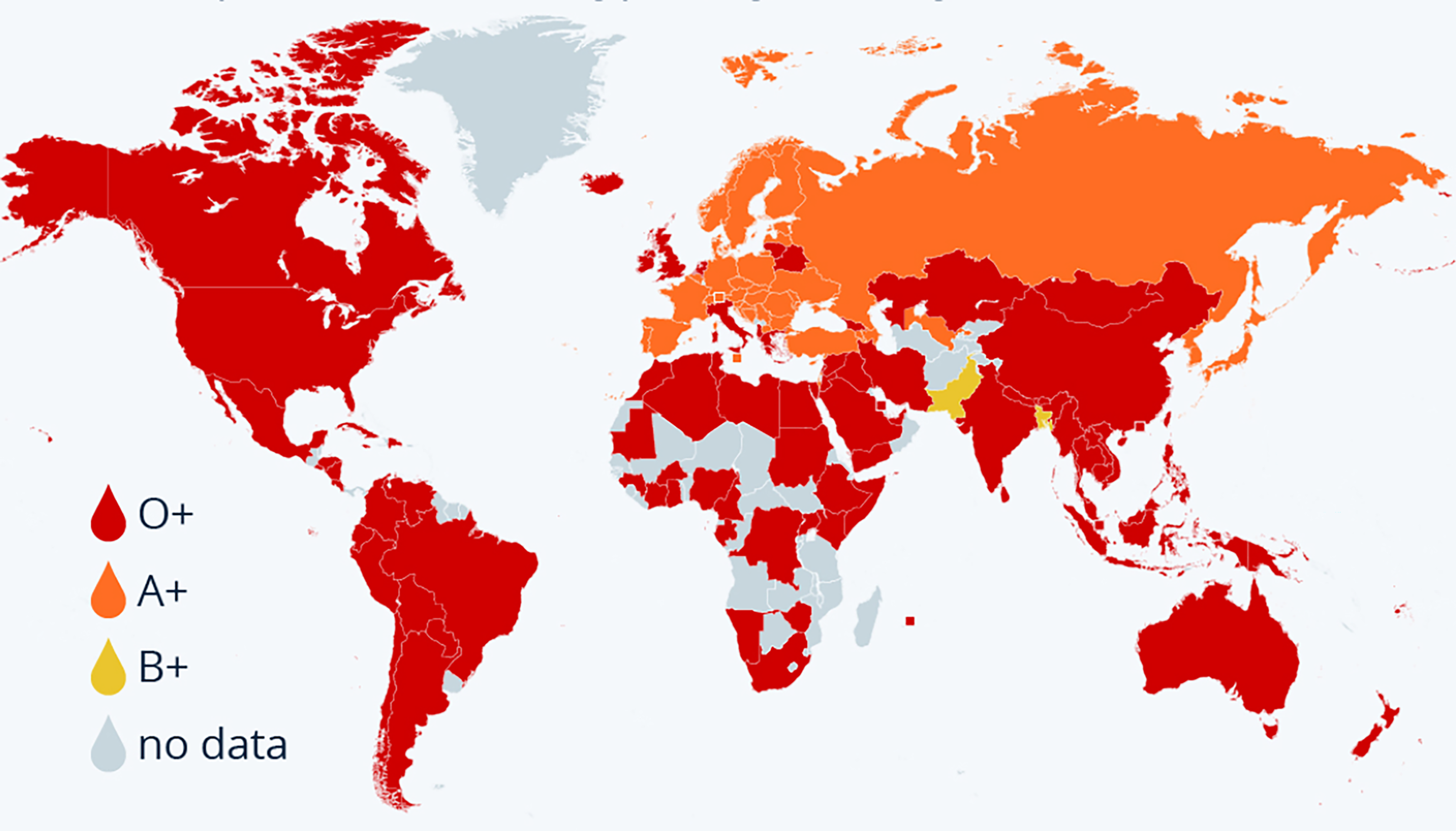
Blood types are found in every human population, but their frequencies vary sharply by region — a pattern shaped by migration, natural selection, and genetic drift in small populations.
The A gene reaches its highest frequencies among some Indigenous Australian peoples, the Blackfoot of Montana, and the Sami of northern Scandinavia. The O gene is common worldwide but especially so among Indigenous peoples of Central and South America. The B gene peaks in Central Asia and northern India. The Rh-negative trait is uncommon in most of the world but reaches notable highs in specific communities: it appears in roughly 15 percent of people of European descent, and highest of all among the Basques of the Pyrenees, at an estimated 25 to 35 percent. Selection leaves its fingerprints too — the Duffy-negative blood that resists vivax malaria is common precisely where that malaria has long been endemic.
The single most common blood type in the world is O positive, carried by an estimated 37 to 39 percent of all people. Grouping by ABO alone, type O leads globally at roughly 39 percent, followed by A at about 31 percent, B at about 24 percent, and AB — the rarest — at about 6 percent. Because the Rh-positive trait is nearly universal (around 85 to 94 percent of people worldwide), the “positive” types dominate everywhere, and AB negative is the rarest of the eight main types, at well under 1 percent.
Japan is the great exception: A positive, not O positive, is its most common blood type.
The leading type shifts by country, and the pattern tells a story of ancestry and migration. O positive is the most common type across most of the world, including the United States; but a broad belt of South and Central Asia — India, Pakistan, Bangladesh, China — carries an unusually high share of type B, and East Asia leans toward type A, with Japan and South Korea standing out. Japan is the notable exception to the global rule: A positive, not O positive, is its most common type.
| Country | Most common type | Notable feature |
|---|---|---|
| World | O positive (~37–39%) | O+ leads on every continent except parts of Europe and East Asia |
| United States | O positive (~37%) | O+ and A+ together cover about 80% of people |
| India | O positive (~37%) | unusually high B positive (~32%), among the world’s highest |
| China | O positive | very high share of type B, similar to India |
| Japan | A positive | rare exception where A+ outnumbers O+; type AB more common than in most nations |
| Most of Europe | A positive or O positive | many countries nearly tied between the two |
| Pakistan and Bangladesh | B positive | among the only nations where B+ is the single most common type |
Blood Groups at a Glance
- Blood groups are defined by antigens on red cells and antibodies in plasma.
- ABO discovered by Karl Landsteiner, 1901; AB added 1902 by von Decastello and Sturli.
- Landsteiner won the 1930 Nobel Prize in Physiology or Medicine.
- Rh factor discovered 1940 by Landsteiner and Wiener, named for the Rhesus macaque.
- The "O" is from the German ohne, "without" (no A or B antigen).
- Universal red-cell donor: O negative. Universal recipient: AB positive.
- For plasma the rule inverts: AB is the universal plasma donor, O the recipient.
- Antibodies: A has anti-B; B has anti-A; AB has none; O has both.
- Antigens: A has A; B has B; AB has both; O has neither.
- A and B are co-dominant; O is recessive; Rh-positive is dominant.
- The ABO gene is on chromosome 9 (9q34.2), with alleles I-A, I-B and i.
- A and B antigens are built on the H antigen: A adds N-acetylgalactosamine, B adds galactose.
- AB negative is the rarest of the eight main types; O positive is the most common (about 37-40%).
- The Rh factor is the D antigen: present is Rh-positive, absent is Rh-negative.
- Rh disease of the newborn needs an Rh-negative mother and Rh-positive fetus; RhoGAM prevents it.
- Kell's K antigen is highly immunogenic; Duffy-negative blood resists Plasmodium vivax malaria.
- Rarest blood: Rh-null, or "golden blood" (fewer than about 50 people documented).
- Bombay phenotype (Bhende, Bombay, 1952): lacks the H antigen, tests as O, needs a Bombay donor.
- An AB x O cross gives only A or B children (never O or AB).
- Agglutination is the clumping of red cells when an antigen meets its matching antibody.
- Blood typing uses forward grouping (cells vs anti-A/anti-B) and reverse grouping (plasma vs known cells).
- The ABO gene predates humans by over 20 million years; chimpanzees are mostly type A.

We Value Your Opinion Comments